American Pigeon Journal
May 1991, page 24.
An Unusual Eye-blister Condition
by Robert J. Mangile
During the 1970's, R.C. Fitch of Virginia sent a photo of a blue grizzled pigeon displaying what appeared to be a small fluid filled "BB" sized blister at the anterior corner of its right eye (Fig. 1). It appeared to be of no consequence but rather unique and the image stayed in my memory.
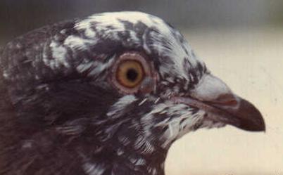
FIGURE 1: A blue/black grizzled pigeon displaying a
small 'blister' near the anterior corner of the right eye.
-- Photo from R.C. Fitch.
Several years later, during a conversation among local fanciers, I described to them my recollection of the photo from Fitch and was surprised to learn that one fancier recalled having a bird in the past with a similar condition. It struck me to be too coincidental and I added his comments to my memory.
On May 23, 1984, an almond-ash-red cock (752-D) was hatched in my
loft which developed a similar blister in the anterior corner of its left
eye. To my best recollection, the blister was first noticed during its
early adulthood. It was a fluid filled 'BB' sized blister and its similarity
to the blue grizzle pigeon in the Fitch photo instantly came to mind.
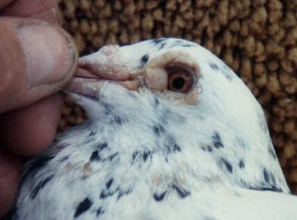
FIGURE 2: An almond-ash-red cock (732-D) displaying a pea-sized
blister on the
anterior corner of the right eye.
-- Photo by Ron Mangile.
These similar cases gave me the impression that they may not be unique and likely occur more frequently than reported. So..., I decided to purposely keep the almond-ash-red cock indefinitely hoping to observe the condition over a long period of time.
Over the past six years he bred and behaved normally and stayed healthy. The eye blister gradually grew larger over the years and seemed to appear more solid, rather than fluid filled. The more solid appearance, I believe, is due to the thickening of the covering skin.
On a few of occasions over the years I've punctured the blister with a needle and later with the tip of a pointed knife, to drain the thick translucent fluid The procedure seemed to inflict little or no pain on the bird and he returned to his normal activities after each operation. The blister always returned!
When photographed on October 16,1990, the blister had grown to approximately pea sized (see Fig. 2). During the early years, the blister posed no problem to the birds vision but it seems to have had some affect during the past couple of years.
Despite the similarity of conditions on the two birds in Figure 1 and Figure 2, their cause may be different. Trying to analyze these conditions seems fruitless; but, it does seem to be a condition that likely occurs more frequently than reported. Perhaps by exposing the pigeon fancy to this uncommon condition, others will come forth with similar stories.
The latter stage of the blister-like development on the almond-ash-red male of mine is gradually beginning to appear like cere and/or wattle tissue. If I were to speculate, my guess would be that it occurs more often in males and in breeds with heavier, rather than lighter, wattles and cere development. Perhaps someone with expertise on the developmental stages of cere and wattle in pigeons may come forth with some helpful comments to enlighten us? Or, someone may offer some other explanation?
# # #

![]()